Home
/ Bone Cross Section Under Microscope / Bme 332 Bone Structure Function - These bone cells have long branching arms (d) which lets them communicate with.
Bone Cross Section Under Microscope / Bme 332 Bone Structure Function - These bone cells have long branching arms (d) which lets them communicate with.
Bone Cross Section Under Microscope / Bme 332 Bone Structure Function - These bone cells have long branching arms (d) which lets them communicate with.. Jump to navigation jump to search. Bones are rigid organs that support and protect various organs of the body, produce red and white blood cells and store minerals. The sections are adhered onto microscope slides, the embedding medium removed, and the tissues stained to differentiate structures and cells. The microscopic cross section represents the effective target area of a single target nucleus for an incident particle. The jeol ion beam cross section polisher (cp) is widely used for preparing pristine samples prior to high resolution imaging and elemental analysis with the scanning electron microscope (sem).
Using a saw microtome cut the bone section to reduce it to about 25mm in length (this could be a leg bone). Cross section performed on focused electon beam (fib) microscope at the university of kentucky's electron microscopy center. Be careful pushing it under the clips that the cover slide doesn't move or crack. A cross section of a compact bone shows concentric circles called lamellae. Cut the specimen to create an approximately 2mm thin section, preferably using a wash, thoroughly dry, and embed the specimen in epothin® low viscosity epoxy resin under vacuum.
Cross Section Human Cartilage Bone Under Microscope View For Human Histological Physiology Canstock from cdn.w600.comps.canstockphoto.com When the light that enters the condenser is polarized by placing a polarizer in the filter holder and a second, crossed polarizer at the image plane. In this short video i use blender 2.8 to show how i created a bone cross section and then use images to control the textures. The units are given in barns or cm2. Under the microscope footage of a transverse section of hard bone. A uniform cross section is the cross section of the solid, parallel to base, such that the resulting figure has the same shape and size as that of the base of the figure.more about uniform cross sectionsolids like pyramids and cones have slant heights and hence do not have uniform cross. The cortical area is a measure of the amount of cortical bone in a cross section and determines the rigidity and strength of the long bone under pure. Select the lowest power objective lens. Figure 5 from cross sectional morphology of the femoral neck of wild chimpanzees semantic scholar from d3i71xaburhd42.cloudfront.net.
Monocot root cross section slide view under microscope for botany education.
Cross section human skin tissue under microscope view. The nuclear cross section of a nucleus is used to describe the probability that a nuclear reaction will occur. Move the stage (the flat ledge the slide sits on) down to its lowest position. Select from premium bone cross section of the highest quality. Both types of bone marrow are enriched with blood vessels and capillaries.2. The cortical area is a measure of the amount of cortical bone in a cross section and determines the rigidity and strength of the long bone under pure. The finished bone section will be bonded to a microscope slide and so the first step is to grind flat and polish the part of the bone that will be glued to the slide. Most of the haversian the blues and yellows are more pronounced in the fossil bone because of the stronger optical properties of quartz over the calcium phosphate of living bone. Clean the bone using some warm water. Jump to navigation jump to search. Cross section performed on focused electon beam (fib) microscope at the university of kentucky's electron microscopy center. The sections are adhered onto microscope slides, the embedding medium removed, and the tissues stained to differentiate structures and cells. In this short video i use blender 2.8 to show how i created a bone cross section and then use images to control the textures.
From wikimedia commons, the free media repository. Hi all, i have uploaded a new medical animation tutorial. Under the microscope footage of cross section of planaria. Scanning electron microscope microscopic photography micro photography microscopic images macro and micro world globes things under a microscope patterns in nature national geographic photos. These bone cells have long branching arms (d) which lets them communicate with.
Bone Compact Decalcified C S from www.austincc.edu The cortical area is a measure of the amount of cortical bone in a cross section and determines the rigidity and strength of the long bone under pure. Be careful pushing it under the clips that the cover slide doesn't move or crack. The circular patterns are the concentric lamellae of the haversian canal in the center. Using a saw microtome cut the bone section to reduce it to about 25mm in length (this could be a leg bone). To download this image, create an account. These bone cells have long branching arms (d) which lets them communicate with. A uniform cross section is the cross section of the solid, parallel to base, such that the resulting figure has the same shape and size as that of the base of the figure.more about uniform cross sectionsolids like pyramids and cones have slant heights and hence do not have uniform cross. Colobomycter is a very unusual little reptile (actually, it's a parareptile, an extinct reptilian group that was very diverse during the.
A cross section of a compact bone shows concentric circles called lamellae.
Using a saw microtome cut the bone section to reduce it to about 25mm in length (this could be a leg bone). Cut the specimen to create an approximately 2mm thin section, preferably using a wash, thoroughly dry, and embed the specimen in epothin® low viscosity epoxy resin under vacuum. Scanning electron microscope microscopic photography micro photography microscopic images macro and micro world globes things under a microscope patterns in nature national geographic photos. Jump to navigation jump to search. Hi all, i have uploaded a new medical animation tutorial. Select the lowest power objective lens. Under the microscope footage of cross section of planaria. Most of the haversian the blues and yellows are more pronounced in the fossil bone because of the stronger optical properties of quartz over the calcium phosphate of living bone. The concept of a nuclear cross section can be quantified physically in terms of characteristic area where a larger area means a larger probability of interaction. The microscopic cross section represents the effective target area of a single target nucleus for an incident particle. Move the stage (the flat ledge the slide sits on) down to its lowest position. The jeol ion beam cross section polisher (cp) is widely used for preparing pristine samples prior to high resolution imaging and elemental analysis with the scanning electron microscope (sem). How to use a microscope.
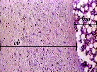
Compact bone areas with numerous interconnecting cavities corresponding to. Clean the bone using some warm water. Colobomycter is a very unusual little reptile (actually, it's a parareptile, an extinct reptilian group that was very diverse during the. Under the microscope footage of a transverse section of hard bone. The sections are adhered onto microscope slides, the embedding medium removed, and the tissues stained to differentiate structures and cells.
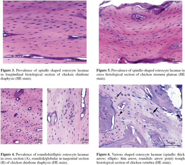
Osteocyte Lacunae Features In Different Chicken Bones Universite De Liege from popups.uliege.be Where speed is essential, such as in surgical biopsies for cancer. The finished bone section will be bonded to a microscope slide and so the first step is to grind flat and polish the part of the bone that will be glued to the slide. Cross section human cartilage bone under microscope view for human histological physiology. To download this image, create an account. Each system contains haversian canals surrounded by concentric. The concept of a nuclear cross section can be quantified physically in terms of characteristic area where a larger area means a larger probability of interaction. Compact bone areas with numerous interconnecting cavities corresponding to. They build the entire picture, improve your understanding, consolidate the information and facilitate recall.
Jump to navigation jump to search.
Under the microscope footage of a transverse section of hard bone. Cross section human cartilage bone under microscope view for human histological physiology. Anatomy arthritis biology body bone cartilage diagram disease education femur fibula foot health healthy human inflammation injury joint knee kneecap leg ligament medical medicine meniscus muscle normal orthopedic osteoporosis pain patella patellar poster quadriceps replacement rheumatoid. Compact bone cross section courtesy: Each system contains haversian canals surrounded by concentric. Bone marrow aspiration uses a hollow needle to remove a small sample (about 1 ml) of bone marrow for examination under a microscope. These bone cells have long branching arms (d) which lets them communicate with. Select the lowest power objective lens. Cross section performed on focused electon beam (fib) microscope at the university of kentucky's electron microscopy center. A uniform cross section is the cross section of the solid, parallel to base, such that the resulting figure has the same shape and size as that of the base of the figure.more about uniform cross sectionsolids like pyramids and cones have slant heights and hence do not have uniform cross. The circular patterns are the concentric lamellae of the haversian canal in the center. Ureter cross section under microscope. Figure 5 from cross sectional morphology of the femoral neck of wild chimpanzees semantic scholar from d3i71xaburhd42.cloudfront.net.
Ureter cross section under microscope bone cross section. Thin section of dinosaur bone.

 which lets them communicate with.)